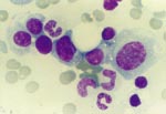
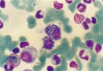
Hemolytic anemia, Leishmania donovani infection, bone marrow plasmatocytosis


Α 58 year-old Greek male was admitted to our Department because of fever, pancytopenia and splenomegaly. Fever, up to 38.5 °C, started fifteen days ago, during the evening. There were no chilling or other symptoms. The administration of amoxycillin for five days had no benefit. Fever continued for the whole day and fatigue, weakness and dyspnea on slight exertion were added. Clarithromycin and cefuroxime were administered for another six days with no effect and the patient was admitted to the hospital.
The physical examination revealed pallor, a cervical microlymphadenopathy and severe splenomegaly (9 cm below costal margin). His temperature was 38.3 °C, the blood pressure was 165/70 mmHg and the pulse rate was 112/min. There was no hepatomegaly or signs of an infection of the respiratory tract and the neurological examination was unremarkable. The patient’s hematology was as follows: white cell count 2.3x10⁹/L (the differential count was: neutrophils 34%, lymphocytes 39% and monocytes 27%), hemoglobin 8.3 g/dL, hematocrit 24.8% and platelet count 88x109/L. The anemia was normocytic and red cell morphology revealed microspherocytes and polychromatophilia. The morphology of white cells and platelets in the peripheral blood smears was normal. The reticulocyte count was 210x109/L and the sedimentation rate was 72 mm/1st hour. The biochemical profile of the patient showed a slight increase of serum transaminases (SGOT 48 U/L, SGPT 52 U/L), a greater increase of bilirubin (2.3 mg/dL; the unconjugated was 1.9 mg/dL) and of lactate dehydrogenase (540 U/L) and severe hypergammaglobulinemia (6.5 g/dL) without a monoclonal component. The quantitative analysis of γ-globulins was as follows: IgG 4.1 g/dL, IgA 1.6 g/dL and IgM 700 mg/dL. The direct Coombs reaction was slightly positive and the red cells were found to be agglutinated with anti-complement serum. Serum haptoglobins were reduced (0.1 g/dL) while the urobilinogen content of the urine was increased. The blood and urine cultures were negative for bacteria or fungi. Tests for HIV, HBV or HCV infection and the Mantoux test were negative. The laboratory investigation for rheumatic diseases, including RA-test, ANA, anti-DNA and anti-ENA antibodies, was negative. There were also no anti-platelet antibodies. CT scan of the thorax revealed mediastinal microlymphadenopathy (the max diameter of the lymph nodes was 1 cm) and CT scan of the abdomen showed splenomegaly. A bone marrow aspiration was performed and a hyperplastic erythroid series was observed. The number of mature plasma cells was increased (at a proportion of about 5–6%) as well as the number of reticule cells.
The intravascular antibiotic treatment had no effect in reducing fever and other symptoms. When the results for the detection of the cause of this febrile splenomegaly were completed the appropriate medication was administered and the patient is now in excellent condition.

Υπενθύμιση στοιχείων εισόδου
  Email
 
  Κωδικός


Νέος χρήστης; Εγγραφή εδώ!