|
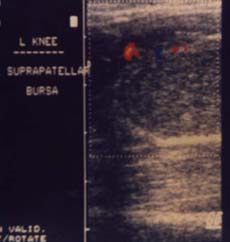
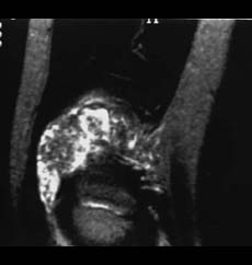
Juvenile chronic arthritis (pauciarticular type of onset) A 2 year old girl in good clinical condition presented with swelling of the right knee. Clinical examination of the knee revealed limitation of extension and flexion. There was no history of trauma or infection. The WBC count was normal, ESR 40 mm, CRP 11.5 mg/dL (normal value<5 mg/dL) with positive ANA. Color Doppler sonography of the knee (left) showed a distended suprapatellar recess filled with echogenic material, and increased vascularity of the synovium. A sagittal T2-weighted MR image of the same knee (right) demonstrated increased signal intensity of the synovium and effusion with some signals of low intensity within the fluid. The inflamed synovium is visualized by increased vasscularity on Color Doppler and high signal intensity on T2- weighted MR images. The echogenic material on US and the low intensity signals on T2- weighted MR images correspond with the "rice bodies" proven by arthroscopy. |